This article was co-authored by wikiHow staff writer, Janice Tieperman. Janice is a professional and creative writer who has worked at wikiHow since 2019. With both a B.A. and M.A. in English from East Stroudsburg University, she has a passion for writing a wide variety of content for anyone and everyone. In her free time, you can find her working on a new crochet pattern, listening to true crime podcasts, or tackling a new creative writing project.
There are 10 references cited in this article, which can be found at the bottom of the page.
This article has been viewed 63,912 times.
Learn more...
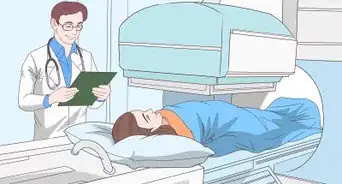
Analyzing a chest x-ray can be a bit of a balancing act. When the patient is rotated to the left or right during the x-ray, the image can look misleading and lead to an incorrect diagnosis. Don’t worry—you don’t need to order a new x-ray if your current one is rotated. However, knowing that your chest x-ray is rotated can help you draw more accurate conclusions and make a decision that’s best for your patient. We’ve outlined a few pointers and tips to help get you on your way.
Steps
Method 1
Method 1 of 11:Collarbone Comparison
-
1Check that the left and right collarbone are equal in length. Ideally, both collarbones should be totally symmetrical. If 1 collarbone looks longer than the other, your x-ray is probably rotated.[1]

Method 2
Method 2 of 11:Collarbone and Spinal Measurement
-
1Measure between the collarbone and central spine. Draw a line down the center of the upper spine, between the throat and the top of the lungs. Then, measure between each collarbone and this middle line. If 1 collarbone is significantly closer or farther to this central line than the other, your x-ray film is likely rotated.[2]

Method 3
Method 3 of 11:Spine and Chest Wall
-
1Measure between the spine and both outer chest walls. The “chest wall” is a fancy term for the left and right sides of the torso. In a rotated x-ray, your spine might look like it’s closer to the left or right side of your chest.[3]

Method 4
Method 4 of 11:Upper Spine
-
1Check that the upper spine is straight and vertical. If the upper spine looks diagonal or uneven, your x-ray could be rotated.[4]

Method 5
Method 5 of 11:Heart Size
-
1Examine the heart to see if it looks especially large or small. When the patient is rotated too much to the left, the heart might look bigger than it actually is. However, if the patient is rotated to the right, their heart might look a bit small. If the heart size doesn’t seem accurate or consistent, there’s a good chance that the chest x-ray is rotated.[5]

Method 6
Method 6 of 11:Soft Tissue
-
1Look for thick sections of soft tissue. When the soft tissue around the chest, like the breast tissue, looks extra thick, doctors might assume the patient has some kind of lung disease. If this happens with your x-ray, there’s a chance that the image could be rotated.[6]

Method 7
Method 7 of 11:Rib Cage
-
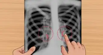
1Check that the left and right rib cages are even in size. If 1 rib cage looks larger or more prominent than the other, the chest x-ray might be rotated.[7]
- Some radiologists designed a method that can accurately measure and analyze rib positioning in chest x-rays. You can find more information here: https://lhncbc.nlm.nih.gov/LHC-publications/PDF/pub8970.pdf

Method 8
Method 8 of 11:Normal Asymmetry
-
1Even if there’s some asymmetry, the x-ray might not be rotated. For instance, the hilar points, diaphragm, and trachea will not always look symmetrical. Here are normal placements for those structures:[8]
- The left lung hilar point is normally higher than the right. However, both should be similar in size and density.[9]
- The right side of the diaphragm is normally higher than the left. The liver sits inferior (below) the right hemidiaphragm.[10]
- The trachea might be slightly to the right (or centered). If it is slightly to the side, be sure to check whether it’s due to pathology.

Method 9
Method 9 of 11:Other X-Rays
-
1Cross-check other x-rays to see if everything adds up. It can be hard to get the full picture from a single x-ray. Before jumping to any conclusions, compare the chest x-ray to any other images that were submitted. These can help you see the full picture, instead of just what the chest x-ray is showing.[11]

Method 10
Method 10 of 11:Inspiration
-
1Like rotation, inspiration also affects how accurate your x-ray will be. Ideally, chest x-rays are taken at the maximum degree of inspiration (inhalation). In a good x-ray, you should be able to see 8-9 posterior ribs. A film taken with poor inspiration can make the structures in the chest look crowded, leading to the appearance of congestive heart failure.[12]

Method 11
Method 11 of 11:Penetration
-
1In addition to lack of rotation, a good x-ray will also have adequate penetration. Penetration (also called exposure) describes how deeply x-rays passed through the body. In a properly exposed chest x-ray, the vertebrae are visible behind the heart. If the film is under penetrated, you won’t be able to see the left hemidiaphragm and can’t assess the lung tissue behind the heart.[13]


References
- ↑ https://iem-student.org/how-to-read-chest-x-rays/
- ↑ https://www.saem.org/cdem/education/online-education/m3-curriculum/group-diagnostic-testing/radiographic-interpretation/chest-radiograph
- ↑ https://www.texaschildrens.org/sites/default/files/uploads/documents/TCHAPP/2019/Chest%20X-ray%20Interpretation-%20The%20ABC%27s.pdf
- ↑ https://www.radiologymasterclass.co.uk/tutorials/chest/chest_quality/chest_xray_quality_rotation
- ↑ https://www.radiologymasterclass.co.uk/tutorials/chest/chest_quality/chest_xray_quality_rotation
- ↑ https://www.radiologymasterclass.co.uk/tutorials/chest/chest_quality/chest_xray_quality_rotation
- ↑ https://lluch.org/sites/lluch.org/files/docs/NICU/2018-CH-Transport-Conference-6N-Mordue-Shedding-Light-on-Neonatal-X-rays.pdf
- ↑ https://www.physio-pedia.com/Chest_X-Rays
- ↑ https://www.radiologymasterclass.co.uk/tutorials/chest/chest_home_anatomy/chest_anatomy_page2
- ↑ https://www.radiologymasterclass.co.uk/tutorials/chest/chest_home_anatomy/chest_anatomy_page7
- ↑ https://www.saem.org/cdem/education/online-education/m3-curriculum/group-diagnostic-testing/radiographic-interpretation/chest-radiograph
- ↑ https://www.youtube.com/watch?v=DNNd_j9hpV0&t=34s
- ↑ https://www.radiologymasterclass.co.uk/tutorials/chest/chest_quality/chest_xray_quality_penetration
- ↑ https://www.saem.org/cdem/education/online-education/m3-curriculum/group-diagnostic-testing/radiographic-interpretation/chest-radiograph
About This Article
Medical Disclaimer
The content of this article is not intended to be a substitute for professional medical advice, examination, diagnosis, or treatment. You should always contact your doctor or other qualified healthcare professional before starting, changing, or stopping any kind of health treatment.